39 microscope with labels and functions
› b › Sony-PlayStation-5-ConsolesSony PlayStation 5 Consoles for sale | eBay Get the best deals on Sony PlayStation 5 Consoles and upgrade your gaming setup with a new gaming console. Find the lowest prices at eBay.com. Fast & Free shipping on many items! recorder.butlercountyohio.org › search_records › subdivisionWelcome to Butler County Recorders Office Copy and paste this code into your website. Your Link Name
Microscope Labels and Functions Flashcards | Quizlet Microscope Labels and Functions. STUDY. PLAY. Ocular Lenses (eyepiece) magnifies. Arm. supports the tube and connects it to the base. Revolving nosepiece. holds the objective lenses and can be rotated to easily change the power. Objective lenses. magnifies. Coarse adjustment knob.

Microscope with labels and functions
en.wikipedia.org › wiki › Two-photon_excitationTwo-photon excitation microscopy - Wikipedia In scattering tissue, on the other hand, the superior optical sectioning and light detection capabilities of the two-photon microscope result in better performance. Applications Main. Two-photon microscopy has been involved with numerous fields including: physiology, neurobiology, embryology and tissue engineering. Compound Microscope Parts - Labeled Diagram and their Functions The eyepiece (or ocular lens) is the lens part at the top of a microscope that the viewer looks through. The standard eyepiece has a magnification of 10x. You may exchange with an optional eyepiece ranging from 5x - 30x. [In this figure] The structure inside an eyepiece. The current design of the eyepiece is no longer a single convex lens. Microscope Parts, Function, & Labeled Diagram - slidingmotion A microscope is a laboratory instrument used to examine very small or micro-objects such as cells and microorganisms that are not able seen by the naked eye. What are the parts of the Microscope? Head Arm Base Eyepiece Lens Eyepiece Tube Objective Lenses Microscope Illuminator Stage and Stage Clips Microscope Nosepiece Rack Stop Condenser Lens
Microscope with labels and functions. en.wikipedia.org › wiki › Electron_microscopeElectron microscope - Wikipedia An electron microscope is a microscope that uses a beam of accelerated electrons as a source of illumination. As the wavelength of an electron can be up to 100,000 times shorter than that of visible light photons , electron microscopes have a higher resolving power than light microscopes and can reveal the structure of smaller objects. Parts of the Microscope with Labeling (also Free Printouts) Parts of the Microscope with Labeling (also Free Printouts) By Editorial Team March 7, 2022 A microscope is one of the invaluable tools in the laboratory setting. It is used to observe things that cannot be seen by the naked eye. Table of Contents 1. Eyepiece 2. Body tube/Head 3. Turret/Nose piece 4. Objective lenses 5. Knobs (fine and coarse) 6. ZEISS Axioscan 7 Microscope Slide Scanner Digitize your specimens with Axioscan 7 – the reliable, reproducible way to create high-quality virtual microscope slides. Axioscan 7 combines qualities that you would not expect to get in a slide scanner: high speed digitization and outstanding image quality plus an unrivaled variety of imaging modes are all available in a fully automated and easy to operate system. Maturation of active zone assembly by Drosophila Bruchpilot Jul 07, 2009 · This led to a complete loss of BRP Nc82 /BRP N-Term labels and T-bars, whereas some traces of residual electron-dense material appeared at the same frequency ... fitted with a 2D Gaussian function with Mathematica (version 5.0; Wolfram Research). The peaks of the Gaussian functions were used to automatically align and subsequently average the ...
abberior LIVE - @abberior.rocks 2-color STED image of a living cell expressing actin K118TAG incorporating TCO*A labeled with abberior LIVE 550 click (gray). In addition, the cell is expressing a SNAP-tag® in an outer mitochondrial membrane protein visualized by our abberior LIVE 610 SNAP (red). Microscope, Microscope Parts, Labeled Diagram, and Functions Microscopes magnify or enlarge small objects such as cells, microbes, bacteria, viruses, microorganisms etc. at a viewable scale for examination and analysis. Microscopes consist of one or more magnification lenses to enlarge the image of the microscopic objects placed in the focal plane. Microscope Types (with labeled diagrams) and Functions This is an advanced microscope that has specific application in viewing, observing and measuring the optical thickness and phase of completely transparent specimens and objects. A tiny interferometer is used and a specimen is placed on beam path of it. This path is split and then rejoined to create two superimposed images of the specimen in focus. Label the microscope — Science Learning Hub Jun 08, 2018 · All microscopes share features in common. In this interactive, you can label the different parts of a microscope. Use this with the Microscope parts activity to help students identify and label the main parts of a microscope and then describe their functions.. Drag and drop the text labels onto the microscope diagram. If you want to redo an answer, click on the …
Microscope Parts & Functions - AmScope Microscope Parts and Functions Invented by a Dutch spectacle maker in the late 16th century, compound light microscopes use two sets of lenses to magnify images for study and observation. The first set of lenses are the oculars, or eyepieces, that the viewer looks into; the second set of lenses are the objectives, which are closest to the specimen. Your liver is essential to your life. The Canadian Liver Foundation This is a myth. Jaundice can be an early warning sign of liver disease. Many babies have “newborn jaundice” lasting three to five days after birth because their liver is not yet fully developed, however, jaundice that does not clear up after 14 days of life, dark urine and/or pale stools, an enlarged abdomen and vomiting are signs that your baby should be seen by his or … Conserved cell types with divergent features in human versus … Aug 21, 2019 · RNA-sequencing analysis of cells in the human cortex enabled identification of diverse cell types, revealing well-conserved architecture and homologous cell types as well as extensive differences ... Binocular Microscope Anatomy - Parts and Functions with a Labeled ... The main function of this aperture of the condenser is to control the light coming up through the condenser. The microscope condenser from a light cone with a sufficiently large angle. And it fills the entire back focal plane of the objective lens. The function of the aperture iris is the same of the photographic lens.
Compound Light Microscope
Microscope labels and functions Flashcards | Quizlet Start studying Microscope labels and functions. Learn vocabulary, terms, and more with flashcards, games, and other study tools.

Biology 2201
Microscope: Parts Of A Microscope With Functions And Labeled Diagram. Q. Define a Microscope. Ans. Microscopes are instruments that are used in science laboratories, to visualize very minute objects such as cells, and microorganisms, giving a contrasting image, that is magnified. Q. State functions of a microscope. Ans. A microscope is usually used for the study of microscopic algae, fungi, and biological specimens.
![Untitled Document [www.stolaf.edu]](http://www.stolaf.edu/people/giannini/cell/ER/rough.gif)
Untitled Document [www.stolaf.edu]
afn.netAmerican Family News Aug 02, 2022 · American Family News (formerly One News Now) offers news on current events from an evangelical Christian perspective. Our experienced journalists want to glorify God in what we do.

Motor Neuron Labeled - Top Label Maker
Microscope labeling and functions Flashcards | Quizlet Microscope labeling and functions STUDY Flashcards Learn Write Spell Test PLAY Match Gravity Created by mveet Terms in this set (27) Separates the eyepiece lens from the objective lenses Body Tube Holds the low-power and high-power objective lenses; allows the lenses to rotate for viewing Revolving Nosepiece Magnifies about 4x
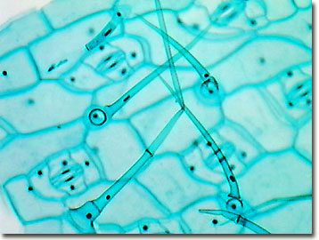
Molecular Expressions: Science, Optics & You - Olympus MIC-D: Brightfield Gallery - Spiderwort ...
Microscope Parts and Functions First, the purpose of a microscope is to magnify a small object or to magnify the fine details of a larger object in order to examine minute specimens that cannot be seen by the naked eye. Here are the important compound microscope parts... Eyepiece: The lens the viewer looks through to see the specimen.
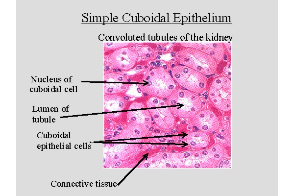
Epithelial Tissue : Anatomy & Physiology
en.wikipedia.org › wiki › CoatingCoating - Wikipedia Functions of coatings. Adhesive – adhesive tape, pressure-sensitive labels, iron-on fabric; Changing adhesion properties Non-stick PTFE coated- cooking pans; Release coatings for example silicone-coated release liners for many self-adhesive products; primers encourage subsequent coatings to adhere well (also sometimes have anti-corrosive ...

32 Compound Light Microscope Label - Labels 2021
A Study of the Microscope and its Functions With a Labeled Diagram ... Electron Microscopes: These illuminate objects with a beam of highly charged electrons. e.g. Transmission electron microscope (TEM) and scanning electron microscope (SEM). These microscopes provide better magnification than light microscopes. Compound Microscope Diagram The compound microscope uses light for illumination.
(22).jpg)
An Ultimate Quiz On Microscope Parts And Functions! - ProProfs Quiz
PharmaCircle This website uses cookies to help provide you with the best possible online experience. Please read our Terms & Conditions and Privacy Policy for information about ...
Post a Comment for "39 microscope with labels and functions"