43 knee joint with labels
› Organic-Hemp-Pain-Relief-Cream › dpHemp Cream - Relieve Muscle, Joint, Back, Knee - Natural Hemp ... This item Hemp Cream - Relieve Muscle, Joint, Back, Knee - Natural Hemp Oil Extract Gel Rub with MSM - Glucosamine - Arnica - Turmeric - Maximum Strength - Made in USA - 4 fl oz Extra Strength Hemp Cream Pain Relief Rub – Only 3rd Party Tested Product To Verify Strength/Results. 3D Knee Joint Model *Finished Product* - YouTube Finally completed my knee joint model with labels of all the key ligaments, muscles, tendons, and bursae. Let me know what you think, I spent a lot of time ...
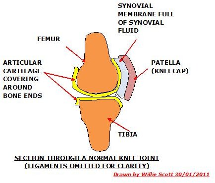
Knee (Human Anatomy): Function, Parts, Conditions, Treatments The knee is one of the largest and most complex joints in the body. The knee joins the thigh bone (femur) to the shin bone (tibia). The smaller bone that runs alongside the tibia (fibula) and the...
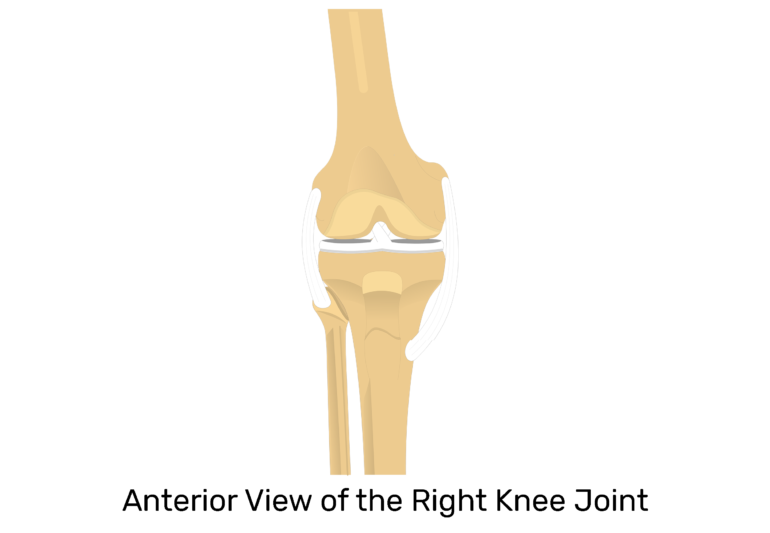
Knee joint with labels
Label The Structures Of The Knee. Chegg - Solved Match The ... Structure of the knee joint 1. Ty label the structures of the knee joint (superior view by clicking and dragging the labels to the correct location lateral menis eu synovial . Label the structures of the knee. Label the structures of the knee. Tibia patellar surface lateral condyle of femur 16 medial condyle of femur anterior cruciate . 🔺 Voodoo Floss How To 💪: knee pain Showing posts with label knee pain. Show all posts. knee knee pain lower limb. Floss Bands for Knee Joint. RockFloss Tutorial on Knee Flossing with Compression Bands Here we'll detail the steps to applying Rockfloss to the knee. The goal will ... Anatomy of human knee joint with labels — text, bones ... "Anatomy of human knee joint with labels" is an authentic stock image by StocktrekImages. It's available in the following resolutions: 1049 x 1600px, 1704 x 2600px, 3422 x 5220px. The minimum price for an image is 49$. Image in the highest quality is 3422 x 5220px, 300 dpi, and costs 449$. Similar Images Same Series Keywords Text Bones
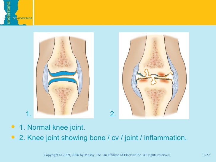
Knee joint with labels. › QUINEAR-Compression-ArthritisAmazon.com: QUINEAR Knee Massager with Heat, Air Compression ... About this item 【Relieve Joint Pain and Help for Injury Recovery】- QUINEAR knee massager has 2+2 airbags inflated and deflated, simulate hand kneading,helps to relax knee and the surrounding calf and thigh area muscles, reduce joint pain and muscle stiffness,the heating will further relieve muscle soreness and help with better circulation, reduce joint swelling, speed up injury recovery. › articles › 310399Osteoarthritis knee pain: Foods to eat and avoid Jan 06, 2022 · Osteoarthritis (OA) of the knee damages the cartilage in the knee joint. Cartilage is a tissue that acts as a cushion at the ends of bones within joints. This results in pain and mobility problems. Knee Anatomy: Bones, Muscles, Tendons, and Ligaments Bones Around the Knee There are three important bones that come together at the knee joint: The tibia (shin bone) The femur (thigh bone) The patella (kneecap) A fourth bone, the fibula, is located just next to the tibia and knee joint, and can play an important role in some knee conditions. Knee Joint - Anatomy Pictures and Information The knee, also known as the tibiofemoral joint, is a synovial hinge joint formed between three bones: the femur, tibia, and patella. Two rounded, convex processes (known as condyles) on the distal end of the femur meet two rounded, concave condyles at the proximal end of the tibia. Continue Scrolling To Read More Below... Additional Resources
Amazon.com: anatomical model knee Axis Scientific Functional Knee Model - Anatomically Correct Knee Joint with Life Like Ligaments That Can Show Movement, Includes Base, Detailed Full Color Product Manual, Worry Free 3 Year Warranty 22 $49 99 Get it as soon as Wed, Apr 13 FREE Shipping by Amazon Labeling the Knee Joint Quiz - PurposeGames.com This is an online quiz called Labeling the Knee Joint There is a printable worksheet available for download here so you can take the quiz with pen and paper. Your Skills & Rank Total Points 0 Get started! Today's Rank -- 0 Today 's Points One of us! Game Points 11 You need to get 100% to score the 11 points available Actions A Labeled Diagram of the Knee With an Insight into Its ... Labeled Diagram of the Knee Joint Knee joint is one of the most important hinge joints of our body. Its complexity and its efficiency is the best example of God's creation. The anatomy of the knee consists of bones, muscles, nerves, cartilages, tendons and ligaments. All these parts combine and work together. Label The Structures Of The Knee Joint - Solved Procedure ... Start studying knee joint label. Man body anatomical poster with bones of. The knee joint is essentially made up of three bones: The thigh bone (femur), the shinbone (tibia), and the kneecap (patella). The knee joint labeled diagram. Label the structures of the knee joint (anterior view) by clicking and dragging the labels to the correct location.
Solved Correctly label the following anatomical features ... Question: Correctly label the following anatomical features of the knee joint. Patellar ligament Synovial membrane Articular capsule Articular cartilage Fat pad Joint cavity This problem has been solved! See the answer Show transcribed image text Expert Answer 100% (1 rating) Articular capsule. Articular … View the full answer Knee x-ray - labeling questions | Radiology Case ... Normal X-ray Knee - Frontal (with labels) Annotated image Frontal Knee Frontal 1. Femoral shaft 2. Patella 3. Base of patella 4. Apex of patella 5. Adductor tubercle of femur 6. Medial epicondyle of femur 7. Medial condyle of femur 8. Lateral epicondyle of femur 9. Lateral condyle of femur 10. Groove for popliteus 11. Intercondylar fossa 12. Knee Joint Anatomy: Structure, Function & Injuries - Knee ... Knee joint anatomy involves looking at each of the different structures in and around the knee. The knee joint is the largest and one of the most complex joints in the human body. There are various muscles that control movement, ligaments that give stability, special cartilage to absorb pressure and various other structures to ensure smooth ... Knee Anatomical Models | Knee Joint Models Knee Models. Because the knee supports almost all of one's body weight, this joint is highly susceptible to injury. With the help of knee models, however, it is easy to see how the three main components, the femur, patella, and tibia, work together, and how they can buckle under stressful situations. Knee education models are useful classroom ...
Lower Extremity Joints Flashcards | Easy Notecards
Knee Joint - San Diego Mesa College Knee Joint. Click on a photo for a larger view of the model. Click on L abel for the labeled model. Back to Muscular System. Anterior: Anterior without patella: Posterior: Label: Label: Label : Label: Label: Label ...

Alila Medical Media | Knee joint diagram unlabeled | Medical illustration
Amazon.com: Wellden Product Anatomical Human Knee Joint ... This life size functional joint model of the right knee consists of the portion of the femur, tibia and fibula as well as the meniscus and patella with the quadriceps tendon and joint ligaments. It shows detailed anatomical structures of the knee joint along with the mechanics of the knee. Size: Life Size Measurement: 11 x 11 x 30cm Weight: 0.5kg
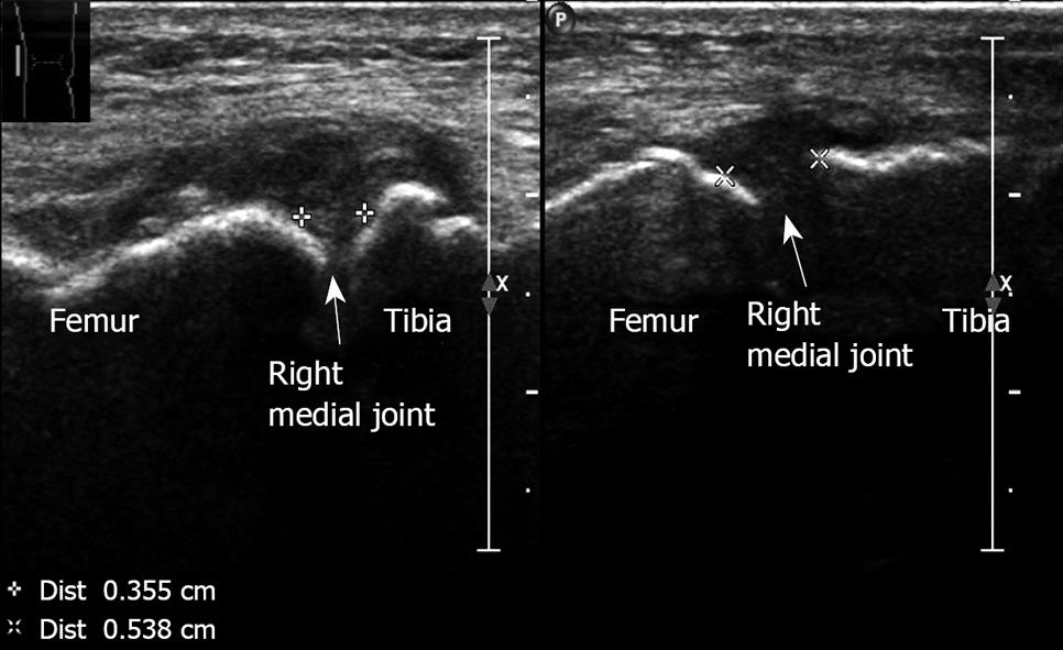
Ultrasound in the diagnosis of clinical orthopedics: The orthopedic stethoscope
Knee Diagram - Pro Knee Pain Relief The knee is a complex joint that has many functions. It bends back and forth and twists minimally from side to side. The knee connects the thigh bone (femur located in the upper leg) to the shinebone (tibia located in the lower leg.The calf bone (fibula located in the lower leg) connects to the joint, but is not directly affected by the hinge joint action.

Synovial Joint of Left Knee Labeling Quiz
A Diagrammatic Explanation of the Parts of the Human Knee ... Knee actually consists of three bones - femur, tibia and patella. Femur is the thigh bone, tibia is the shin bone and patella is the small cap like structure which rests on the other two bones. Femur is considered as the largest bone in the human body. The femur and the tibia meets at the tibiofemoral joint and patella rests on top of this joint.
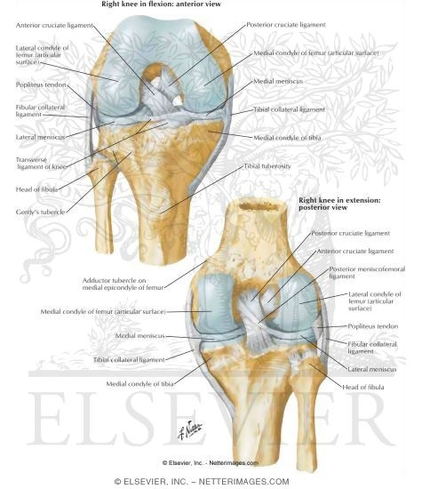
Cruciate and Collateral Ligaments of Right Knee Joint Knee: Cruciate and Collateral Ligaments
Knee Joint Labeled Diagram stock vector. Illustration of ... Knee Joint Labeled Diagram Royalty-Free Stock Photo A knee joint with detailed labels anatomy knee, knee anatomy, joint cartilage, detailed labels, knee, labels, joint, doctor, health, anatomy, medicine, pain, osteoporosis, arthritis, disease, bone, leg, tear, femur, cartilage, disc, shading, acl, ligament, tibia More ID 39627491

Illustration Knee Meniscus Surgery Tendon Graft (Print #8251703)
Knee Joint Anatomy (Labeling) Diagram - Quizlet Only $2.99/month Knee Joint Anatomy (Labeling) STUDY Learn Flashcards Write Spell Test PLAY Match Gravity Created by diegoparas Terms in this set (20) A Femur B Patellar Space Surface C Lateral Condyle D Lateral Collateral Ligament (LCL) E Lateral Meniscus F Transverse Ligament G Fibular Head H Tibial Tuberosity I Medial Condyle J

Alila Medical Media | Knee joint labeled. | Medical illustration
Alila Medical Media | Knee joint, basic labels | Medical ... Human knee joint diagram showing joint cavity, capsule, all cartilage. - Alila Medical Media
Patella Bone - Anterior and Posterior Views
Solved 3 of 5 B. Structure of the knee joint 1. Label the ... Label the parts of the knee joint models anterior cruciate ligament, femur, fibula, fibular collateral ligament, meniscus, patella, patellar ligament, posterior cruciate ligament, tendon of the quadriceps, tibia, tibial collateral ligament 2. Give the functions of the following structures often found in a synovial This problem has been solved!

Knee Joints
Knee Joint Label Flashcards - Quizlet Knee Joint Label STUDY Flashcards Learn Write Spell Test PLAY Match Gravity Created by LaLaKub91 Terms in this set (10) femur What is A? lateral collateral ligament what is d? lateral meniscus what is e? fibula what is g? tibia what is h? posterior cruciate ligament What is j? anterior cruciate ligament what is k? medial meniscus what is l?
Best Exercise Equipment for Arthritic Knees
Knee Joint Picture Image on MedicineNet.com The knee functions to allow movement of the leg and is critical to normal walking. The knee flexes normally to a maximum of 135 degrees and extends to 0 degrees. The bursae, or fluid-filled sacs, serve as gliding surfaces for the tendons to reduce the force of friction as these tendons move. The knee is a weight-bearing joint.
Bone Structure Medical Educational Science Vector Stock Vector 334392599 - Shutterstock
Knee joint: anatomy, ligaments and movements | Kenhub The tibiofemoral joint Medial condyle of femur Condylus medialis femoris 1/7 The tibiofemoral joint is an articulation between the lateral and medial condyles of the distal end of the femur and the tibial plateaus, both of which are covered by a thick layer of hyaline cartilage .

Human Anatomy Lab: Knee Joint Model
› pain › knee-pain-how-toHow to Relieve Knee Pain - Consumer Reports Jun 24, 2018 · A 2015 Cochrane review on bracing found that wearing a brace, such as a valgus knee brace, neutral brace, or neoprene sleeve, may have little or no effect on pain reduction, knee function, or ...

MRI Musculo-Skeletal Section: MRI anatomy of the shoulder (sagittal view).
Knee Anatomy, Diagram & Pictures | Body Maps The knee is the meeting point of the femur (thigh bone) in the upper leg and the tibia (shinbone) in the lower leg. The fibula (calf bone), the other bone in the lower leg, is connected to the...

Human Anatomy Lab: Knee Joint Model
dynref.engr.illinois.edu › amlFour-Bar Linkages - University of Illinois Urbana-Champaign Example: Knee joint (constrained motion) The human knee joint is a type of biological hinge, which allows movement in only one primary angle. The knee connects the femur (the upper leg bone) to the tibia (the larger of the two lower leg bones). These two bones sit next to each other and are free to rotate about a single axis.
M med chapter_001
nurseslabs.com › 5-total-joint-knee-hip5 Total Joint (Knee, Hip) Replacement Nursing Care Plans Mar 18, 2022 · Joint replacements are indicated for irreversibly damaged joints with loss of function and unremitting pain, selected fractures, joint instability and congenital hip disorders. Total Joint Replacement can be performed on any joint except the spine. Hip and knee replacements are the most common procedures.
Post a Comment for "43 knee joint with labels"